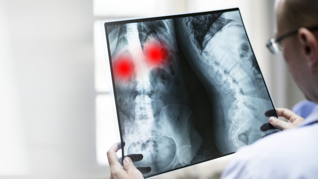
At Novomed, we recognize that imaging procedures are one of the most valuable resources our specialists have to diagnose and advise patients. For this reason, we have an highly experienced French- and UK-trained radiologist heading the department. We offer specialized forms of imaging such as neuroimaging, alongside X-rays, MRIs and CT scans in Dubai.
Our Services Include
- Women’s imaging including hysteroscope
- Breast imaging (mammograms, ultrasound)
- Breast biopsy
- Ultrasound (breasts, thyroid, veins, arteries, etc)
- MRI
- CT scan
- Fluoroscope
- Barium swallows and enemas
- Neuroimaging
- X-rays
What is the Difference Between a CT Scan, MRI and Ultrasound?
A computerized tomography (CT) scan combines a series of X-rays taken from different angles to make detailed picture of a part of your body, for example, organs, blood vessels, bones and the spinal cord. To have the scan, you lie down on a table attached to the scanner, which sends X-rays through the body part being studied. Each scanner rotation takes an image of a thin slice of the body part, and then all of these images are combined and can be printed. The scan is used both for diagnosis and to plan medical, surgical or radiation treatment.
Magnetic resonance imaging (MRI) uses powerful magnets, radio waves, and a computer to take detailed images of a body part such as organs, tissues and skeletal system. Unlike a CT scan, or x-rays, an MRI does not use radiation. The scan can be used both for diagnosis of an illness or injury, and to monitor how well you are responding to treatment.
Ultrasound uses sound waves to create images of a body part. To form an image, an instrument called a transducer sends out high-frequency sound, and then records the echoes as the sound waves bounce back. This helps to determine the size, shape, and consistency of soft tissues and organs. The information is relayed to a computer screen in real time. The image created is used to diagnose conditions and to guide doctors through medical procedures.
Mammograms
During a mammogram, the breast is compressed between two plates and an X-ray is transmitted through the breast tissue. The images that are captured are called mammograms. ‘Screening mammograms’ normally involve two or more x-rays of each breast. The images often make it possible to detect tumors that can’t be felt, and can also find microcalcifications, which sometimes indicate the presence of breast cancer. ‘Diagnostic mammograms’ capture more x-ray images of the breasts in order to obtain views from several angles. The technologist might also magnify a suspicious area to assist the doctor in the diagnosis.
Detecting Stroke Risk With Ultrasound
Ultrasound imaging, also called sonography, is a pain-free and safe way to produce pictures of the inside of the body using sound waves. They can be used to screen arteries to find problems before the symptoms even begin. Carotid arteries are the two main arteries that carry oxygen-rich blood from the heart to the brain. If plaque (a combination of cholesterol, fat and other substances) builds up on the artery walls, it causes athereosclerosis. Over time, the walls of the arteries thicken and narrow, limiting the flow of blood. This causes carotid artery disease, which greatly increases your risk of having a stroke. The goal of ultrasound screening is to detect disease at its earliest and most treatable stage.
When is a Thyroid Ultrasound Done?
Ultrasound imaging, also called sonography, is a pain-free and safe way to produce pictures of the inside of the body using sound waves. Usually, an ultrasound of the thyroid is performed for the following reasons:
- to examine the appearance of nodules on the thyroid and to establish if they require a biopsy
- If a nodule has been felt during a physical exam, the doctor will want to know if there are additional nodules, which an ultrasound can detect
- to monitor whether a nodule has grown substantially over time
- to establish if a lump in the neck is on the thyroid or an adjacent structure
If you are interested in our Radiology and Imaging services, book your consultation today with our experienced Radiologists by calling toll-free 800 (NOVO) 6686 or fill the booking form below.